

PLATEX | ANONYMIZED CASE STUDY
Recovering cardiac action potentials for concentration-dependent assessment.
PlateX helped a research team turn noisy cardiomyocyte assay signals into usable cellular evidence for assessing concentration-dependent changes and selecting follow-up work.
THE SCIENTIFIC QUESTION
The input was a derivative-like or instrument-proxy signal related to membrane potential, rather than a direct action-potential recording. An assay-appropriate, model-informed reconstruction supported assessment of cellular repolarization. Separately measured hERG current remained a distinct source of evidence.
- The challenge
- Noisy proxy recordings related to membrane potential
- The intervention
- Assay-appropriate, model-informed reconstruction
- The outcome
- Concentration-related cellular evidence for follow-up
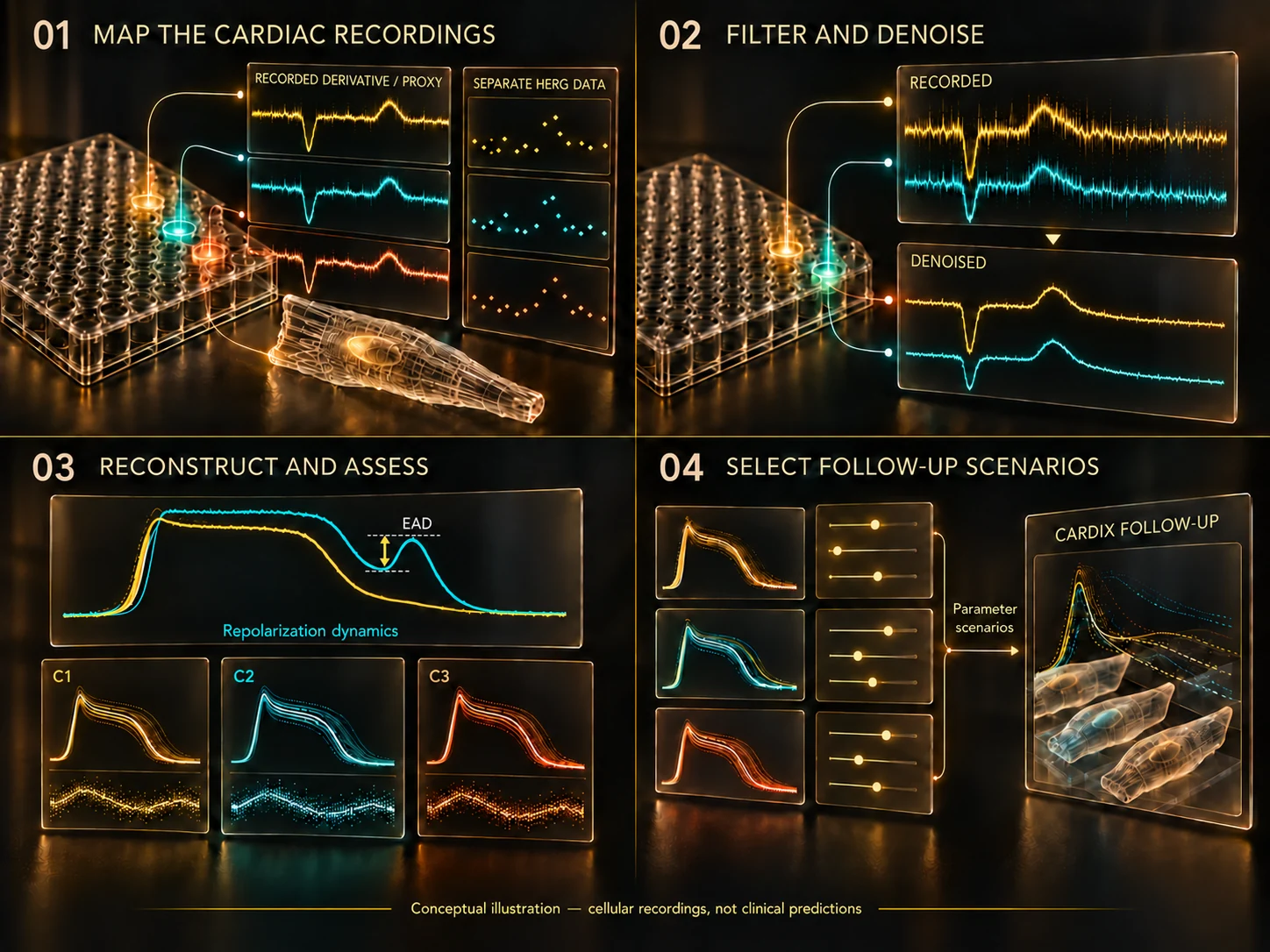
01 / MAP THE EXPERIMENTAL EVIDENCE
Connect each signal to its concentration and context.
The client recorded a noisy proxy for membrane potential in a cardiomyocyte assay. ADETERA mapped these recordings to drug concentrations and the available experimental context, preserving the distinction between the measured proxy and the action potential to be reconstructed.
The separately measured hERG-current dataset retained its own measurement provenance. These channel-level measurements were distinct from the proxy used for waveform reconstruction.
02 / FILTER AND DENOISE
Prepare the recorded proxy for reconstruction.
Substantial noise obscured features needed for concentration-dependent assessment. ADETERA applied filtering and denoising tailored to the recorded signals and the analytical question.
The meaning of the proxy and the assay context determined how the processed signal could support reconstruction. A derivative-like signal was not assumed to be an exact derivative of membrane potential. The mathematical methods and implementation details remain proprietary to ADETERA.
03 / RECONSTRUCT AND ASSESS
Assess action-potential duration and repolarization.
This workflow for cardiac electrophysiology signal analysis used an assay-appropriate, model-informed reconstruction to produce cellular action-potential waveforms. Assessment against the client’s feature specifications examined concentration-response patterns, including action-potential duration (APD) prolongation and repolarization features.
The assessment also considered possible early afterdepolarizations (EADs): secondary depolarizations during repolarization, before the cell has fully returned to its resting state. These were candidate cellular features for interpretation and follow-up, without a clinical risk prediction or safety classification.
04 / DEFINE THE FOLLOW-UP
Turn cellular findings into questions for the next study.
The analysis identified parameter directions for subsequent investigation and a possible handoff to CardiX. Reconstructed waveform features and the separate hERG measurements could inform questions for later simulation while retaining their distinct origins and limitations.
CardiX simulation was outside this case. The work delivered an assessment of concentration-related cellular features and directions for further research.
WHAT THIS ENABLED
Cellular evidence to guide the next study.
The outcome was a structured assessment of concentration-related cellular features from noisy proxy recordings, with directions for further investigation. Any subsequent CardiX simulation would be a separate stage of work.